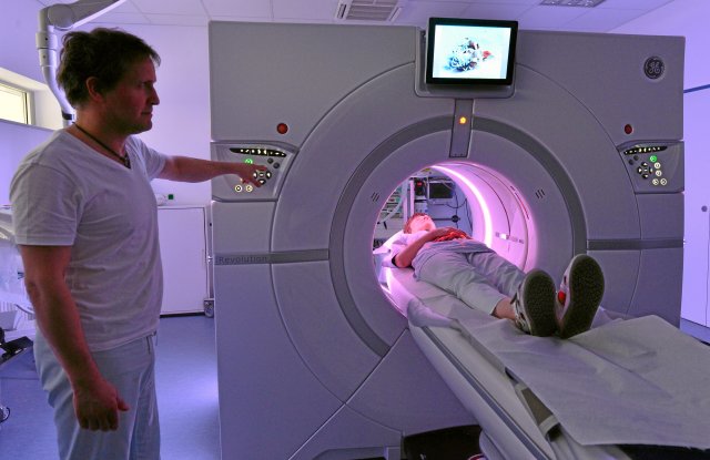
Inconceivable without higher mathematics: diagnoses using computer tomography
Photo: dpa/Martin Schutt
Nowadays, almost every adult has been in the “tube” for a computer tomography (CT) or magnetic resonance imaging (MRI). Or more specialized tomography is carried out. Even though some people find the confines of these devices oppressive, it is becoming increasingly easier to look into every inner corner of the human body. And three-dimensional diagnostic procedures are becoming more and more specific. In January, for example, it was decided that in the future, if people with statutory health insurance were suspected of having chronic coronary heart disease, computed tomography coronary angiography (CCTA) could be used, meaning that a cardiac catheter examination would no longer be necessary.
Conventional X-ray diagnostics, on the other hand, could initially only project the three-dimensional structures onto a two-dimensional surface. The key idea for representation without loss of information is thanks to the mathematician Johann Radon (1887 – 1956). He introduced a new integral transformation of spatial functions – later named after him. The stroke of genius in his paper, which appeared in a volume of the “Saxon Society of Sciences” in 1917, is expressions for the inverse transformation to the Radon transformation. His formulas can be interpreted as instructions for use for certain real processes, for an imaging process later called tomography (from the Greek tomos = section).
nd.DieWoche – our weekly newsletter

With our weekly newsletter nd.DieWoche look at the most important topics of the week and read them Highlights our Saturday edition on Friday. Get your free subscription here.
Cutting into layers
A material body is given; what is sought is the distribution of the mass or selectively certain components in it. To do this, a group of parallel planes is selected to serve as a reference system. The desired distribution in the body is made up of the distributions in the sections with the planes. A beam is then sent through the body from each direction in each reference plane. X-rays or another type of radiation is used. As they pass through the body, these rays are weakened. What then arrives is measured by detectors and stored.
In summary, a data set X is converted into a data set Y; X are the relevant material properties of the body, Y consists of the detector displays. Radon’s transformation describes the causation of Y by X. The most interesting question is: Can one infer the cause of X from the effect of Y? Yes, Radon’s inverse transformation does just the reconstruction! This theoretical result has immense practical consequences: In principle, you can see inside a body with such a tomography. The doctor receives information about the inside of an organism; They are made up of sectional image information and can be accessed in the desired order. Technicians can inspect a workpiece anywhere non-destructively. Archaeologists determine rich details of finds without damaging them.
Long computing times
Of course, it was a long way from the principle to implementation. He began with a discouraging message: the radon inverse transform is poorly conditioned, in other words, error-prone. A small deviation in Y (for example an unavoidable rounding error) can distort the result X. The art of mathematics found a solution to the problem; Roughly speaking, it consists of the interposition of additional integral transformations. However, this exacerbates the next problem: the known calculation methods fail when evaluating a tomography. Formally they work, but they take too long. Even the best computers would be overwhelmed, and even more so would human patience. Mathematicians therefore invented more efficient calculation methods with moderate running times. Physicists were and are required when it comes to materials, radiation and intensities. Finally, engineers build the corresponding large-scale devices.
This is how the Nobel Prize in Medicine was awarded in 1979 to the physicist Allan McCormack and the engineer Godfrey N. Hounsfeld for significant advances in the development of computer-assisted tomography.
Today, in addition to X-rays, other penetrating rays are also used: those from the electromagnetic spectrum, particle radiation (from electrons, positrons or neutrons) and even sound. The material properties sought today are correspondingly diverse. The well-known CT shows the mass distribution, the MRI shows the distribution of the protons in the water content.
Subscribe to the “nd”
Being left is complicated.
We keep track!
With our digital promotional subscription you can read all issues of »nd« digitally (nd.App or nd.Epaper) for little money at home or on the go.
Subscribe now!
